
Without exposing the patient to ionizing radiation, ultrasound can help medical professionals evaluate a wide range of diseases. These may include pain, swelling, abnormal growths, blood flow issues, pregnancy-related concerns, and more.
What is Sonography?
This imaging method has been a crucial component of medical diagnosis, from tracking pregnancy to examining blood flow through veins and arteries to evaluating unexplained stomach pain.
Simply put, it’s a non-invasive medical imaging technique that produces images of organs, tissues, blood vessels, and other structures using high-frequency sound waves.
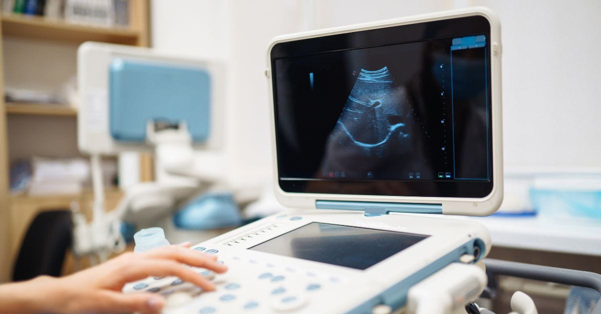
A portable instrument known as a transducer or probe is used for the examination. After sound waves enter the body and interact with various tissues, the probe detects the echoes returning. These echoes are processed by a computer and turned into visuals shown on a monitor.
The device introduces sound into the body, listens for echoes to return, and then uses the data to create an image. This may involve blood flowing through vessels, fetal movement, or organ movement, depending on the investigation.
How Does Sonography Test Work?
Several simple stages are in a standard ultrasonography examination, including:
- In a position that permits access to the area under examination, the patient lies or sits.
- The skin is treated with a transparent ultrasonic gel.
- The gel facilitates the effective transfer of sound waves between the body and the probe.
- The doctor starts to move the probe or transducer across the examination area.
- High-frequency sound waves are transmitted into the body via the probe.
- Different tissues and structures reflect these waves in different ways.
- The returning echoes are processed by the ultrasound system, which then displays images on the screen/ monitor.
Though some pressure from the probe may cause mild discomfort if the area being probed is already tender, the examination is usually painless. Unless their medical team recommends otherwise, most routine scans don't require a recovery period. So, patients can resume their regular activities afterward.
What Is Sonography Used to Diagnose?
Sonography's adaptability is one of the reasons it's so effective. When examining symptoms like discomfort, swelling, a lump, or a possible infection, doctors may advise an ultrasound.
Plus, it can offer real-time guidance during specific treatments or help monitor an existing medical condition.
Typical uses for diagnostics include:
- Abdominal Sonography
An ultrasound of the abdomen can look at structures like:
- The liver
- The gallbladder
- The pancreas
- The kidneys and Spleen
- Blood vessels in the abdomen
When physicians need to look into symptoms like edema, stomach pain, possible gallbladder issues, urinary issues, or other issues, they may recommend it.
- Pelvic Sonography
The uterus and ovaries are among the structures that pelvic ultrasonography can assess. Investigating pelvic pain, irregular bleeding, ovarian abnormalities, or other reproductive-system issues may be helpful, depending on the clinical circumstances.
- Pregnancy Ultrasound
Ultrasound assesses the pregnancy and tracks fetal progress, and it doesn't require ionizing radiation to deliver information in real time.
- Breast Sonography
Besides assessing a breast lump or other anomaly, breast ultrasonography can offer other details about the breast tissue. When necessary, this can also help with image-guided breast biopsies.
- Thyroid and Neck Ultrasound
The thyroid and other soft tissue structures in the neck can be examined with sonography. This can investigate thyroid nodules, swelling, or other anomalies.
- Ultrasound of the Musculoskeletal System
Also, several muscles, tendons, ligaments, and soft tissue structures can be examined using ultrasound. Imaging might occasionally help observe structures during movement because it takes place in real time.
- Heart and Vascular Imaging
Echocardiography also uses ultrasound to look at your heart. And, vascular ultrasonography can evaluate veins and arteries and detect issues such as blood clots or constricted channels.
What Is Doppler Ultrasound?
This is a specific type of ultrasound used to assess blood flow. Doppler techniques give information on the flow of blood through blood vessels rather than just the structure of an organ or tissue.
Doppler ultrasonography helps evaluate:
- Blood flow via veins and arteries
- Constriction or obstruction of blood vessels
- Potential blood clots
- Decreased blood supply to an organ
- Some vascular anomalies
Different Doppler methods exist. Spectral Doppler graphically depicts flow measurements, whereas color Doppler uses colors to show information about blood flow direction and speed.
While power Doppler does not offer the same directional information as color Doppler, it can be especially sensitive when blood flow is relatively low.
Which Sonography Types Are Most Common?
The medical question under investigation determines the type of ultrasonography that is chosen.
- External ultrasound: The skin is touched with the transducer. The most common kind of ultrasound examination is this one.
- Internal ultrasound: To get closer pictures of specific structures, a specially made transducer can be placed inside a bodily hole. The process will be explained in advance by the medical expert.
- Doppler ultrasound: Mainly used to assess blood flow via arteries and movement in general.
- 3D ultrasound: In suitable clinical applications, 3-dimensional representations are produced using gathered ultrasound data.
- Ultrasound-guided procedures: During procedures like biopsies or fluid aspiration, medical professionals can use real-time ultrasound to guide certain needles or tools.
How Should You Prepare for Sonography?
The body part being checked determines your preparation. Certain pelvic exams may need a full bladder, while some ultrasound tests require fasting for a predetermined amount of time. There may be little to no preparation required for a few tests.
Instead of thinking that every ultrasound needs the same preparation, patients should follow guidelines given by their imaging center or doctors. Another way to facilitate the inspection is to dress comfortably and loosely on the day of the ultrasound.
Is Sonography Safe?
The fact that diagnostic ultrasound doesn't use ionizing radiation is one of its most vital advantages. Here, ultrasound uses sound waves, and when used properly for medical purposes, diagnostic ultrasonography is generally safe, according to major medical sources.
Professionals with the necessary training should conduct ultrasounds and interpret the results.
What Happens After the Ultrasound?
A trained medical practitioner, usually a radiologist, reviews the images taken during the examination (ultrasound). The results are recorded in a report and forwarded to the physician who ordered the test.
Then, your doctor will explain the report in relation to your symptoms, medical history, and other test findings. A final diagnosis cannot always be made from an ultrasound scan alone. If more information is needed, your healthcare provider can suggest blood tests, CT, MRI, biopsy, or another study.
What Are the Limitations?
Ultrasound is not the ideal imaging test for every portion of the body, but its versatility. It can be challenging to get clear images of some structures because sound waves cannot pass through air or bone.
Now, based on the patient's body composition and the location being checked, ultrasound may also have trouble imaging very deep tissues. For these reasons, when ultrasonography is unable to provide enough information, clinicians/ doctors may suggest other imaging techniques like CT or MRI.
Final Words
Sonography or ultrasonography has established itself as one of the most useful diagnostic medical instruments. It enables medical professionals to view beneath the skin without the need for surgery or ionizing radiation.
Ultrasound can give insights quickly and safely. If you are looking for sonography in Siliguri under a highly reputed expert, you might connect with Dr. Shubhi Goyal.
FAQs
1. Are sonography and ultrasound the same thing?
They are the same; the terms sonography and ultrasonic imaging typically refer to the same diagnostic imaging method.
2. Is radiation used in ultrasounds?
No, instead of using ionizing radiation, diagnostic ultrasound uses high-frequency sound waves.
3. Does sonography hurt?
Though there may be some pressure or brief discomfort, a sonography test is painless.
4. What is the duration of a sonography test?
Many ultrasound exams take between 30 and 60 minutes, while the exact time varies depending on your advised examination.
5. Can ultrasound detect every disease?
No, ultrasound has limitations, especially when it comes to imaging deep tissue, bone, or air-obscured locations.
6. Who evaluates the ultrasound findings?
The images are interpreted by a licensed radiologist or other suitably trained medical professional. Upon receiving your report, you have to visit your doctor for an explanation.


